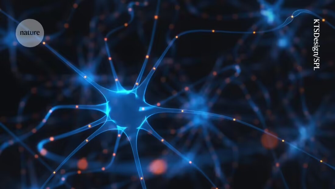
A collection of sticky substance that covers neurons in a Appetite control center in the brain captures, is associated with the deterioration of Diabetes and obesity associated, as a study on mice shows 1.
This substance also prevents insulin from reaching the neurons in the brain that control hunger. Inhibiting the production of this substance led to weight loss in the mice, the experiments found. These results indicate that there is a new trigger for Metabolic disorders that could help scientists identify drug targets to treat these diseases.
These results were published today in the journalNaturepublished.
Hunger regulators in the brain
Metabolic diseases such as Type 2 diabetes and obesity can occur when the body's cells become insensitive to insulin, a hormone that regulates blood sugar levels. Scientists looking for the mechanism that causes this insulin resistance have focused on a part of the brain called arcuate nucleus of the hypothalamus is known. This area detects insulin levels and adjusts energy consumption accordingly the feeling of hunger to.
When the animals developed insulin resistance, a type of... cellular framework, called the extracellular matrix, which holds the hunger neurons in place, into a disorganized substance. Previous research had shown that this framework changes when mice are fed a high-fat diet 2.
The researchers wanted to find out whether these changes in the brain might cause insulin resistance, rather than just occurring at the same time. They fed mice a high-fat, high-sugar diet for 12 weeks and monitored the scaffolding around hunger neurons by taking tissue samples and monitoring gene activity.
They found that this framework became thicker and stickier within a few weeks of starting the unhealthy diet. As the animals gained weight, their hypothalamic neurons became less able to process insulin normally, even when the hormone was injected directly into their brains. This suggests that the stickiness of the scaffold prevents insulin from entering the brain. Instead, “it gets stuck,” says co-author Garron Dodd, a neuroscientist at the University of Melbourne in Australia.
Loss of the substance leads to weight loss
To reverse these changes, the researchers injected the mice with either an enzyme that breaks down the substance or a molecule called fluorosamine that inhibits the formation of the scaffold. Both approaches successfully removed the sticky obstacle in the animals' brains, thereby increasing insulin uptake. Fluorosamine even caused the animals to lose weight and increase their energy expenditure. Treating insulin resistance by targeting the supporting scaffolding around neurons may be safer than targeting the neurons directly, says Dodd.
This "high-quality" study proves "again and again" that this cellular scaffold regulates hormonal signaling, which has direct effects on the body's metabolism and drives disease, says Kimberly Alonge, a biochemist at the University of Washington School of Pharmacy in Seattle, who was not involved in the study. It also draws attention to the need to look not only at individual cells and cell types, but also at the “packaging material in which the cells sit,” she adds.
The team's experiments also showed that inflammation in the hypothalamus drives the disruption of the framework. However, the study does not clarify what originally triggers the inflammation, says Alonge. Previous research has shown that brain cells called glia can influence the structural integrity of the scaffold, and Alonge wants to know whether glial cells contribute to the inflammation in the study.
It remains unclear what role dysfunctional scaffolding plays in the development of metabolic diseases compared to other well-established triggers, says Dodd. He and his colleagues hope to address this question later.
Further research is needed to investigate whether this sticky material arises in people who develop metabolic diseases. This could be challenging, says Dodd, because there is no non-invasive access to the hypothalamus, which lies deep in the brain, and it is difficult to collect tissue samples even from donated organs.

 Suche
Suche
 Mein Konto

